X-Rays
X-rays are types of electromagnetic radiation probably most well-known for their ability to see through a person's skin and reveal images of the bones beneath it. Advances in technology have led to more powerful and focused X-ray beams as well as ever greater applications of these light waves, from imaging teensy biological cells and structural components of materials like cement to killing cancer cells.
X-rays are roughly classified into soft X-rays and hard X-rays. Soft X-rays have relatively short wavelengths of about 10 nanometers (a nanometer is one-billionth of a meter), and so they fall in the range of the electromagnetic (EM) spectrum between ultraviolet (UV) light and gamma-rays. Hard X-rays have wavelengths of about 100 picometers (a picometer is one-trillionth of a meter). These electromagnetic waves occupy the same region of the EM spectrum as gamma-rays. The only difference between them is their source: X-rays are produced by accelerating electrons, whereas gamma-rays are produced by atomic nuclei in one of four nuclear reactions.
Why Are X-Rays Done?
- Broken bones
- Dislocated joints
- Arthritis
- Abdominal pain, in some instances
- Cancer
- Tooth decay
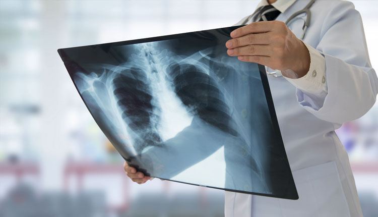
Doctors can also use X-rays to find an object that a child or adult swallowed. An X-ray can be used to check your lungs for signs of pneumonia or tuberculosis, to figure out why you have shortness of breath, or to see if you have heart failure.
Other ways doctors use specific X-ray procedures include:
Mammography: This is an exam that puts your breast between a support plate and a second plate called a paddle, then a series of X-rays are taken. Doctors look closely at the images for signs of cancer or other issues.
Computed tomography (CT) scan: A computer puts together a series of X-rays, taken from different angles, to make a 3D image and give your doctor a more detailed picture.
Fluoroscopy: Sometimes called an ”X-ray movie,” this procedure shoots a continuous X-ray through a part of your body so doctors can see that part and how it moves. It’s most commonly done to look at bones, muscles, joints, and organs like your heart, kidneys, and lungs.